
Happy and Healthy
In Your Own Skin
Understanding the Role of Pathology in Mohs Surgery
Mohs micrographic surgery is renowned as the gold standard for treating many common and high-risk skin cancers. Unlike traditional surgical excisions where only a small percentage of the margins are checked, Mohs surgery utilizes immediate, on-site pathological analysis. This unique feature is the very foundation of the procedure’s success. By combining the roles of surgeon and pathologist, we can precisely map and examine 100% of the tissue margins, guaranteeing complete cancer removal while preserving the maximum amount of surrounding healthy tissue. This commitment to precision is what makes Mohs the most effective treatment available today.
Real-Time Microscopic Mapping and Analysis
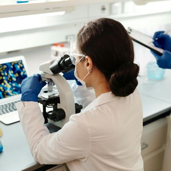
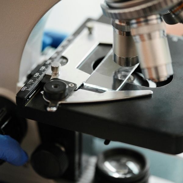
The pathology process is what sets the Mohs procedure apart. Once a layer of tissue is removed, it is immediately taken to our in-house lab, color-coded, and mapped with extreme accuracy. A specialized technician then quickly freezes and slices the tissue horizontally. This allows for microscopic examination of the entire peripheral and deep margins. We evaluate these frozen sections in real-time while the patient waits comfortably. This rapid analysis is critical because it eliminates the uncertainty of standard excision and guides the subsequent surgical steps with unmatched precision. We believe this immediate feedback is essential.
Why 100% Margin Check Matters for Cure
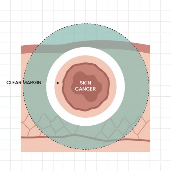
The goal of any cancer surgery is achieving clear margins, meaning no cancer cells remain at the edges of the removed tissue. Standard methods only check small portions of the margins, leaving room for microscopic “roots” of cancer to be missed. With Mohs pathology, we examine the entire margin, virtually eliminating guesswork. If cancer cells are detected, their exact location is mapped. We then remove only the tissue precisely corresponding to that location, sparing healthy tissue elsewhere. This systematic, layer-by-layer approach ensures the highest published cure rates for many skin cancers. For details on our methods, see our Mohs Skin Cancer Surgery page.
The Specialized Role of the Mohs Pathologist
Our surgeon is uniquely trained to act as both the surgeon who removes the tissue and the pathologist who interprets the slides. This dual expertise eliminates delays and communication errors inherent in sending specimens to an outside lab. We possess specialized knowledge in dermatopathology, allowing us to accurately differentiate subtle cancer cells from normal tissue on the frozen sections. This integration of roles ensures that every decision during the procedure is based on the most current and complete microscopic evidence. Our focus is always on the complete eradication of the tumor.
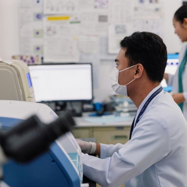
Benefits of In-House Laboratory Processing
Having a dedicated, on-site laboratory is a significant advantage for our patients undergoing Mohs micrographic surgery. It allows us to process and analyze slides quickly, minimizing the time the patient spends waiting between stages. Faster results lead to a quicker, safer procedure and a smaller final wound, which significantly improves the reconstruction and cosmetic outcome. This seamless integration of surgical and pathology teams underscores our commitment to comprehensive, efficient, and precise care right here at our skin cancer surgery center. We aim to maximize both cure and cosmetic results.
Pathology is not merely a step in the Mohs surgery process; it is the cornerstone that guarantees its efficacy. By performing microscopic analysis in real-time, we ensure complete cancer removal with minimal impact on healthy skin. We are proud to offer this meticulous level of care as a leading skin cancer surgery center. If you have questions about the pathology or surgical stages, or if you need to schedule a skin check, please contact Colorado Skin Surgery & Dermatology. We are dedicated to providing precise, compassionate, and comprehensive dermatologic care.